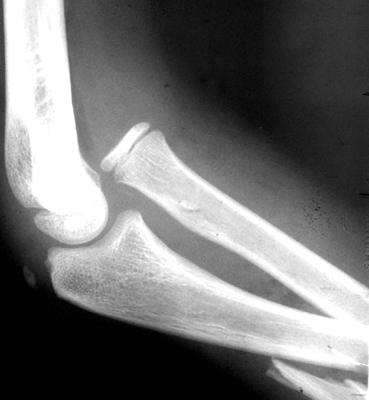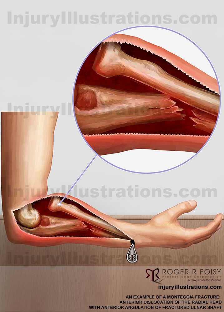

Bado decades ago, involves ulnar fracture and concomitant dislocation of the radial head. Monteggia fracture, named after Giovanni Monteggia in the nineteenth century and well described and classified by Dr. This case, with occult proximal ulna fracture, angulated radial neck fracture, subsequent radiocapitellar dislocation, and articular incongruity, was deemed as a rare Monteggia type-one equivalent fracture-dislocation variant rather than an ordinary radial neck fracture and it facilitates further understanding and management of the Monteggia fracture. And acceptable results were achieved 1 year later.

Then a definitive operation was performed, which involved a Boyd incision, correction of radial head tilting, opening wedge osteotomy of the proximal ulna and proper fixation respectively. A further reduction to the fracture and joint site only resulted in a subluxated and incongruous radiocapitellar joint on the three-dimensional computed tomography (3D-CT). Acceptable results were acquired at first-week follow-up, yet dramatic changes turned up 2 weeks later when the dislocated radial head was found. Case presentationĪ 11-year-old girl, whose injury pattern initially appeared to be a mild radial neck fracture with undisplaced proximal ulnar fracture, and without radial head dislocation, was treated with closed reduction and long-arm splint immobilization. The aim of this study was to present a furthermore type of that lesion which no previous study had reported and arouse pediatric orthopedists’ additional awareness of it. The term has gradually evolved since its introduction, as sporadic case reports continued to complement it.

Please see our publications on radius fractures.Monteggia equivalent lesion represents a series of combined elbow and forearm injuries that resemble typical Monteggia fracture either in presentation or mechanism. The HSS Orthopedic Trauma Service has conducted many studies. Lateral radiograph of the left-sided Monteggia fracture-dislocation respectively.Īnteroposterior and lateral radiographs at 12 months illustrating healed Monteggiaįracture-dislocations in excellent alignment.Ĭlinical pictures at 12 months demonstrating excellent range of motion.

Kloen P, Rubel IF, Farley TD, Weiland AJ, Helfet DL: "Bilateral Monteggia fractures."Īnteroposterior, and lateral injury radiographs of the right-sided Monteggia fracture-dislocation and She most recently returned for routine follow-up at 1 year following fracture surgery with excellent radiographic and clinical results including healed Monteggia fractures, complete resolution of pain, excellent range of motion, and she has returned to all activities of daily living. Weiland with placement of plates, screws a single cerclage wire (left-side) and interfragmentary lag screws. Weiland, MD at the Orthopedic Trauma Service at Hospital for Special Surgery. Radiographs revealed bilateral (right- and left-sided) Monteggia fractures with posterior dislocations of both radial heads (Bado type-II injuries). Case Example Bilateral Monteggia Fracture DislocationsĪ previously healthy female, 53 years of age, fell when she tripped while carrying a laundry basket down steps landing on the dorsal aspects of her flexed elbows, and had immediate pain in both upper extremities.


 0 kommentar(er)
